Recherche translationnelle en santé,
technologie pour la santé et recherche clinique

iDREAM team / Axis 1 : Imaging of brain motor repair
Multi-modal Imaging of regenerative medecine:
Brain implants and cellular therapy
The axis is led by Isabelle Loubinoux and Franck Desmoulin.
Scientific objectives
We develop therapies for patients with severe disabilities (stroke, severe traumatic brain injury).
Our approach is based on regenerating brain lesions with implants and/or cell transplants.
MRI imaging is a crucial tool for longitudinal monitoring to assess the integration of implants or cells in the brain, their vadcularization, the degradation of biomaterials, their safety and security, as well as the detection of adverse effects.
Regenerative implants
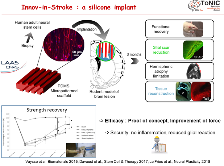
The functionalized silicone implant
Regenerative medecine offers new hope for treatment based on neuro-implants (cell therapy using biomaterials) to restore long-distance connections between brain areas. The team has demonstrated proof of concept for neuro-implants that improve motor performance in rodents. Safety has been demonstrated in non-human primates (marmosets).
A clinical trial of implant safety is being prepared: the FAVOUR project (ANR Regenerative Medicine and Messidore programs) on patients with severe head trauma.
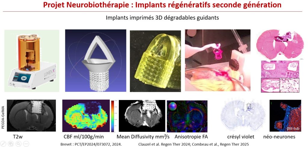
The 3D printed degradable implant
The second generation of regenrative implants focuses on biodegradable implants. 3D printing gives them a complex architecture mimicking the cerebral cortex in six interconnected layers. T2-weighted imaging provides good visualization of the hydrogel and its degradation over time. pCASL perfusion imaging allows monitoring of implant revascularization. Diffusion tensor imaging measures the diffusivity of water within the material, pores, and tissue (Matrix project, ANR ASTRID funding).
Cellular therapy
Intranasal graft of enteric glial cells
The proximity of enteric nerve cells to brain cells could make them a source of backup for the brain (Colitti et al. Adv Sci 2026).
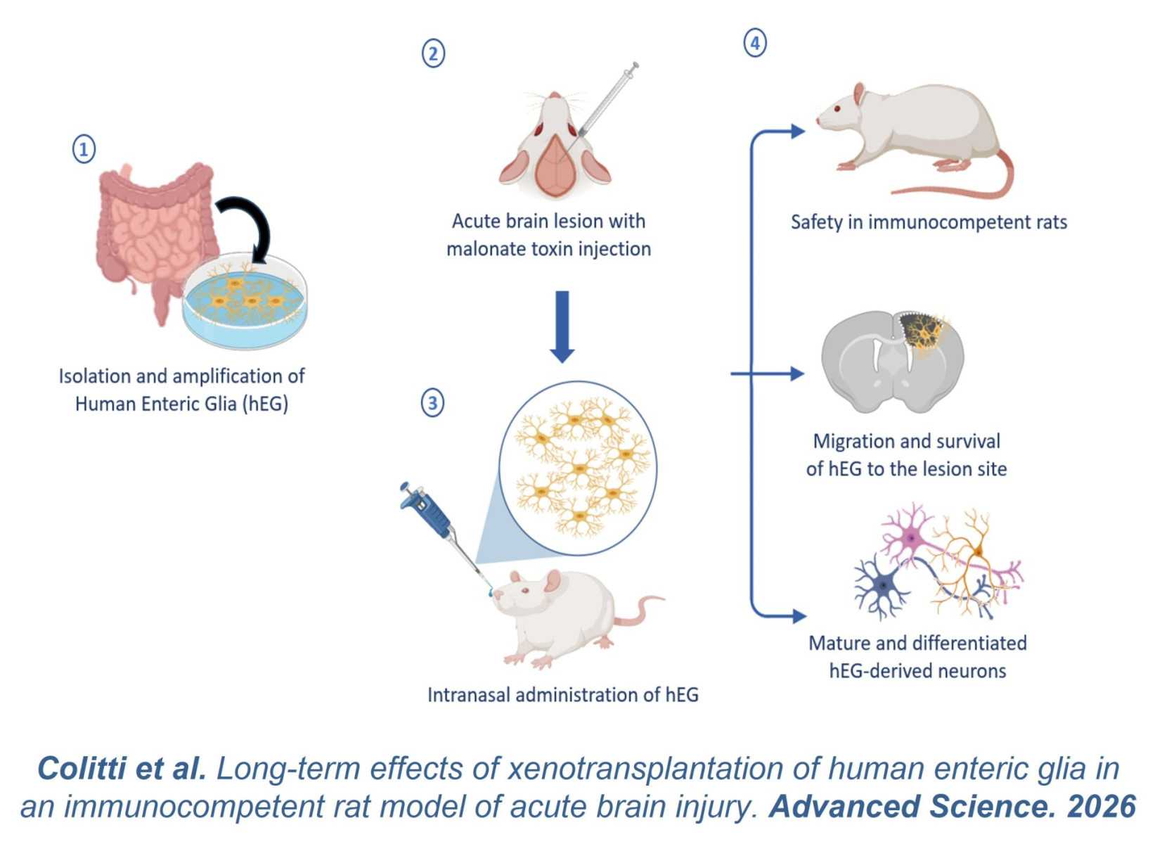
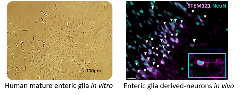
Human enteric glial cells were intranasally grafted into rat brains and preferentially differentiated into neurons (Colitti et al. Adv Sci 2026).
Preclinical imaging
Secondary degeneration biomarker
We have discovered a new biomarker of gliosis accompanying axonal degeneration in stroke patients and in brain-lesioned non-human primates (marmoset). For several months after the cortical lesion, the corticospinal tract undergoes a loss of myelin, an astrocytic infiltration and a microglial reaction. They induce a loss of intensity on T2 MRI scans and a slight hyperintensity on T1 MRI scans (Le Friec et al., Trans Stroke Res 2020).
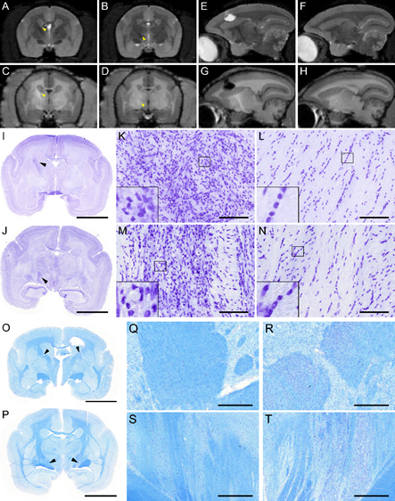
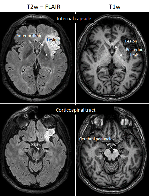
Evidence of corticospinal tract degeneration
Opening of the blood brain barrier
Our brain is protected by the blood-brain barrier. But some drugs cannot get through it.
Preclinical imaging is used to develop procedures for temporary opening of the blood-brain barrier, to determine safe conditions and cerebral blood flow modifications.
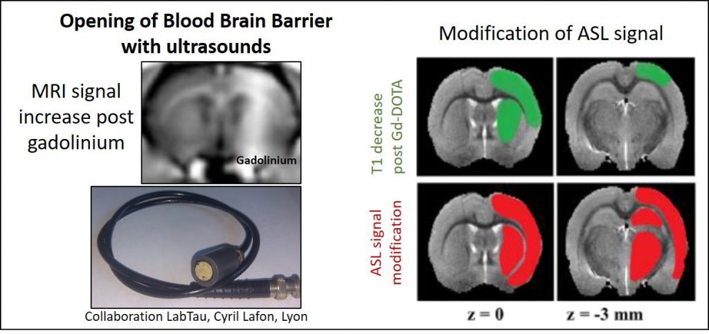
(Labriji et al. Magnetic Resonance in Medecine, 2023)