Recherche translationnelle en santé,
technologie pour la santé et recherche clinique

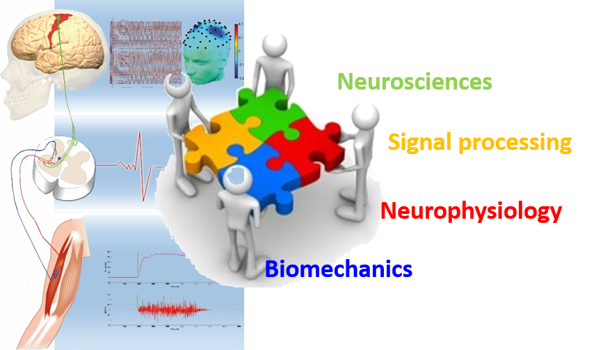
iDREAM team / Axis 2 : Neurobiomechanics


![]()
Neurobiomechanical – Biomarkers of physiologic and pathologic motricity
The objective of the work developed in this line of research is to provide a better understanding of the nervous mechanisms involved in the control of movement and motor learning.
Through a multidisciplinary approach at the interfaces between fundamental, clinical and translational research, we explore cognitive and motor functions and the underlying spinal and supra-spinal nervous mechanisms involved in the control of healthy or altered muscle contractions in healthy individuals, experts and neurological patients.
This axis led by David AMARANTINI et David GASQ.
Our research activities deal with the following four topics :
— Topic 1 : Spinal and supraspinal nervous mechanisms involved in the muscle activity control.
— Topic 2 : The role of rhythm in motor learning and rehabilitation for children with motor disorders
— Topic 3 : Multidimensional approach to spastic co-contractions in brain damaged subjects.
— Topic 4 : Nervous mechanisms involved in the emotional flexibility of motor control.
Team members
Permanent members
Associated members
Alexandre CHALARD – ![]()
![]()
PhD students
Master 2 students
Vincent ARDONCEAU – ![]()
Anaïs DESBERNATS – ![]()
Thibaut GELY – ![]()
Pierre GOUDY
Emmeline MONTANE – ![]()
Morgane PATERNOSTER
Anastasia THEODOSIADOU – ![]()
Lisa VICTORIA – ![]()
Collaborations

Clinical imaging
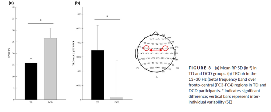
Coordination disorders and Electroencephalography
Jessica Tallet and co-workers recorded EEG in children with development coordination disorders (DCD) and controls with typical development (TD). They demonstrated that atypical inter-hemispheric communication correlates with altered motor inhibition during learning of a new bimanual coordination pattern in developmental coordination disorder. Indeed, appropriate motor control involves inhibition of mirror movements, which relies on inter-hemispheric connectivity and coherence (RP: relative phase; TRCoh: task-related coherence) (Blais et al., Developmental Science, 2018).
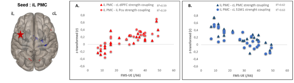
Stroke and MRI functional connectivity
MRI functional connectivity during a sensitivomotor task was investigated in 25 stroke patients and was correlated with motor deficit (Fugl-Meyer Scale of upper limb, FMS-UE). Negative connectivity of ipsilesional premotor cortex with contralesional motor regions (blue clusters) is observed in very impaired patients and evidences disconnection. A cross-modal functional connectivity of the ipsilesional premotor cortex with non-motor regions (prefrontal cortex, precuneus) ws evidenced and may reflect efficient cross-modal compensation strategies (red clusters) (Brihmat et al., Brain Connectivity 2020).
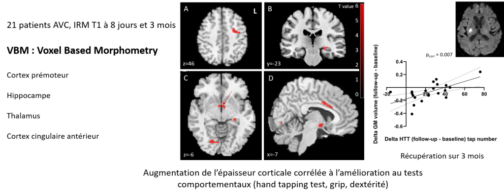
Stroke and cerebral plasticity with multimodal MRI
Brain plasticity was explored in 21 patients with capsular lesions and pure motor deficits at 4 days (SD 3) post-stroke and at 4 months. The fractional anisotropy of the corticospinal tract (FACST) was significantly reduced on the affected side at 4 months and correlated with motor performance (9-peg hole test, dynamometer, tapping test). FACST is therefore a good biomarker for diagnosing post-stroke motor deficits. Voxel-based morphometry (VBM) revealed increases in cortical thickness in several regions, which correlated with performance. The presence of alternative motor tracts was not correlated with recovery. In this group of patients with mild deficits, it would appear that good recovery relies on preserved corticospinal fibers, particularly the ipsilesional premotor cortex. VBM and FACST are thought to be reliable biomarkers of atrophy, reorganization, plasticity, and post-stroke recovery (Loubinoux et al., J Neurol 2024).